Study of Renal Tubular Cell Damage by Oxidative Stress in Potassium Deficient Rats(การศึกษาการทำลายเซลล์เยื่อบุผนังท่อไตเนื่องจากกระบวนการออกซิเดชั่นในภาวะขาดโพแทสเซียมในหนูแรต)
Keywords:
Potassium depletion(ภาวะพร่องโพแทสเซียม), Oxidative stress(ภาวะออกซิเดชั่น), Renal tubular injury(การบาดเจ็บของท่อไต)Abstract
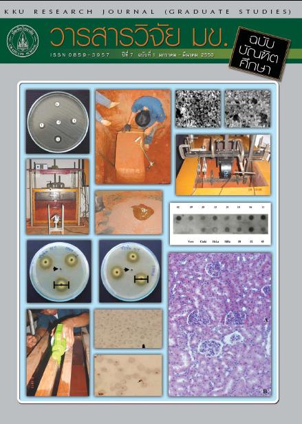
Our aim was to study the effect of potassium (K+ ) depletion (KD) on oxidative damage of renal tubular cells. Male Sprague - Dawley rats were fed three periods with normal diet (control group) and K+ deficient diet for 7, 14 and 21 days (KD group). After blood and 24-hour urine samples were collected at the end of each period, rats were scarified to obtain kidneys and soleus muscle. Plasma and muscle were analyzed for K+ content and kidney for reactive oxygen species (ROS) and superoxide dismutase (SOD; both CuZnSOD and MnSOD) activity and urine for N-acetyl-β-glucosaminidase (NAG). Histological pattern changes of the kidneys were also examined. The results showed that K+ contents of KD group were significantly decreased in both plasma and muscle. While the ROS formation and MnSOD activity were significantly increased (p<0.05), the Cu Zn SOD activity was unchanged in the KD group. The NAG activity in urine of the KD group was significantly increased (p<0.05). These changes were clearly observed since after the 7 day of feeding. Changes in histological appearance were less interstitial lesion, diffuse epithelial cell swelling and vacuolar degeneration. Our results indicate potassium depletion can cause renal tubular epithelial oxidative damage especially after 14 and 21 days of feeding .
การศึกษานี้มีวัตถุประสงค์เพื่อดูผลของภาวะพร่องโพแทสเซียมที่มีต่อการทำลายเซลล์เยื่อบุผนังท่อไตเนื่องจากกระบวนการออกซิเดชัน ทำการเลี้ยงหนูแรตพันธุ์ Sprague-Dawley เพศผู้ด้วยอาหารธรรมดา (กลุ่มควบคุม) และอาหารขาดโพแทสเซียม (กลุ่ม KD) เป็นเวลา 7, 14 และ 21 วัน หลังจากเก็บตัวอย่างเลือดและปัสสาวะ 24 ชั่วโมง ในแต่ละช่วงของการให้อาหารทำการฆ่าหนูเพื่อเก็บตัวอย่างกล้ามเนื้อลายชนิด soleus กับไต ทำการวิเคราะห์หาปริมาณโพแทสเซียมในพลาสมาและกล้ามเนื้อลาย ปริมาณสารอนุมูลอิสระของออกซิเจน (ROS) และการทำงานของเอนไซม์ superoxide dismutase (SOD) ทั้งชนิด CuZnSOD และ MnSOD) ในไตและวัดอัตราการทำงานของเอนไซม์ N-acetyl--glucosaminidase (NAG) ในตัวอย่างปัสสาวะรวมทั้งดูลักษณะการเปลี่ยนแปลงของเนื้อเยื่อไต ผลการศึกษาพบว่าระดับโพแทสเซียมในทั้งของพลาสมาและกล้ามเนื้อของหนูกลุ่ม KD ลดลงอย่างมีนัยสำคัญ