Biochemical Responses and DNA Damage of Chlorella pyrenoidosa H. Chick upon Exposure to Combined Cu and Cd at Environmentally Realistic Levels DOI: 10.32526/ennrj.18.3.2020.28
Main Article Content
Abstract
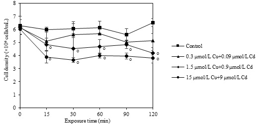
Monitoring aquatic ecosystems is necessary to prevent or reduce the impact of metal pollutants on ecosystems and human health. Biological responses, or biomarkers, can provide quick and direct evidence of exposure to environmental stressors. This study evaluated the sensitivity of biomarkers of Chlorella pyrenoidosa after short-term exposure to combined copper and cadmium at environmentally realistic concentrations with the following biological endpoints: growth; alkaline phosphatase activity; chlorophylls, pheophytin-a, carbohydrate, and protein content; and DNA damage. C. pyrenoidosa was exposed to three combinations of copper and cadmium for 120 min, or for 48 h to assess its DNA damage. To assess the sensitivity of the biological responses to the combined metals, integrated biomarker response (IBR) analysis was also performed. The results demonstrated that exposure to combined Cu and Cd caused an inhibition of growth and activity of alkaline phosphatase, a decrease of chlorophyll-a and -b and protein, and an increase of pheophytin-a and tail factors DNA. The IBR analysis affirmed that the inhibition of alkaline phosphatase activity and the decrease of protein level were responsive biomarkers for exposure to the combined metals.
Article Details
Published articles are under the copyright of the Environment and Natural Resources Journal effective when the article is accepted for publication thus granting Environment and Natural Resources Journal all rights for the work so that both parties may be protected from the consequences of unauthorized use. Partially or totally publication of an article elsewhere is possible only after the consent from the editors.
References
2. Arunakumara KKIU, Xuecheng Z. Heavy metal bioaccumulation and toxicity with special reference to microalgae. Journal of Ocean University of China 2008;7(1):60-4.
3. Awasthi M. Relevance of alkaline phosphatase activity of immobilized green algae and cyanobacteria for heavy metal toxicity monitoring. Journal of Materials and Environmental Science 2012;3(3):446-51.
4. Bačkor M, Váczi P. Copper tolerance in the lichen photobiont Trebouxia erici (Chlorophyta). Environmental and Experimental Botany 2002;48:11-20.
5. Bajguz A. Suppression of Chlorella vulgaris growth by cadmium, lead, and copper stress and its restoration by endogenous brassinolide. Archives of Environmental Contamination and Toxicology 2011;60:406-16.
6. Bartell SM. Biomarkers, bioindicators, and ecological risk assessment: A brief review and evaluation. Environmental Bioindicator 2006;1:60-73.
7. Bertrand L, Monferrán MV, Mouneyrac C, Bonansea RI, Asis R, Amé MV. Sensitive biomarker responses of the shrimp Palaemonetes argentinus exposed to chlorpyrifos at environmental concentrations: Roles of alpha-tocopherol and metallothioneins. Aquatic Toxicology 2016;179:72-81.
8. Cadet J, Wagner JR. DNA base damage by reactive oxygen species, oxidizing agents, and UV radiation. Cold Spring Harbor Perpectives in Biology 2013;5(2):1-18.
9. Campanella F, Cubadda F, Sammartino MP, Saoncella A. An algal biosensor for the monitoring of water toxicity in estuarine environments. Water Research 2000;35(1):69-76.
10. Cheng J, Qiu H, Chang Z, Jiang Z, Yin W. The effect of cadmium on the growth and antioxidant response for freshwater algae Chlorella vulgaris. Springerplus 2016;5(1)1290:1-8.
11. Cid A, Herrero C, Torres E, Abalde J. Copper toxicity on the marine microalga Phaeodactylum tricornutum: Effects on photosynthesis and related parameters. Aquatic Toxicology 1995;31:165-74.
12. Desai SR, Verlecar XN, Nagarajappa, Goswami U. Genotoxicity of cadmium in marine diatom Chaetoceros tenuissimus using the alkaline comet assay. Ecotoxicology 2006;15(4):359-63.
13. Elfidasari D, Ismi LN, Sugoro I. Heavy metal contamination of Ciliwung River, Indonesia. Ecology and Safety 2019;13:106-11.
14. Erbes M, Weβler A, Obst U, Wild A. Detection of primary DNA damage in Chlamydomonas reinhardtii by means of modified microgel electrophoresis. Environmental and Molecular Mutagenesis 1997;30:448-58.
15. Ferro Y, Durrieu C. Development of an algal biosensor for toxicity assessment of urban wet weather effluents. Proceedings of the SIDISA 2012 Sustainable Technology for Environmental Protection; 2012 Jun 26-29; Politecnico di Milano, Milan: Italy; 2012.
16. Grigoryev Y. Cell counting with a hemocytometer: easy as 1, 2, 3 [Internet]. 2014 [Cited 2020 Jan 18]. Available from: http://bitesizebio.com/13687/cell-counting-with-a-hemocytometer-easy-as-1-2-3/.
17. Gani P, Sunar NM, Matias-Peralta H, Latiff AAA, Razak ARA. Influence of initial cell concentrations on the growth rate and biomass productivity of microalgae in domestic wastewater. Applied Ecology and Environmental Research 2016; 14(2):399-409.
18. Hazlina AZ, Devanthiran L, Fatimah H. Morphological changes and DNA damage in Chlorella vulgaris (UMT-M1) induced by Hg2+. Malaysian Applied Biology 2019;48(1):27-33.
19. Ihlenfeldt MJA, Gibson J. Phosphate utilization and alkaline phosphatase activity in Anacystis nidulans (Synechococcus). Archives of Microbiology 1975;102:23-8.
20. Iturburu FG, Bertrand L, Mendieta JR, Amé MV, Menone ML. An integrated biomarker response study explains more than the sum of the parts: Oxidative stress in the fish Australoheros facetus exposed to imidacloprid. Ecological Indicators 2018;93:351-7.
21. Ivancsits S, Diem E, Pilger A, Rudiger HW, Jahn O. Induction of DNA strand breaks by intermittent exposure to extremely low frequency electromagnetic fields in human diploid fibroblasts. Mutation Research 2002;519:1-13.
22. Jiang H, Islam MdS, Sazawa K, Hata N, Taguchi S, Nakamura S, Sugawara K, Kuramitz H. Development of an electrochemical bioassay based on the alkaline phosphatase activity of Chlamydomonas reinhardtii to assess the toxicity of heavy metals. International Journal of Electrochemical Science 2016;11:5090-102.
23. Kruger NJ. The Bradford method for protein quantitation. In: Walker JM, editor. Methods in Molecular Biology: Basic Protein and Peptide Protocols Vol. 32. New Jersey: Humana Press, Inc.; 1994. p. 9-15.
24. Küpper H, Küpper F, Spiller M. In situ detection of heavy metal substituted chlorophylls in water plants. Photosynthesis Research 1998;58:123-33.
25. Lecoeur S, Videmann B, Berny PH. Evaluation of metallothionein as a biomarker of single and combined Cd/Cu exposure in Dreissena polymorpha. Environmental Research 2004; 94:184-91.
26. Leonard SS, Harris GK, Shi X. Metal-induced oxidative stress and signal transduction. Free Radical Biology and Medicine 2004;37:1921-42.
27. Levy J, Angel B, Stauber JL, Poon WL, Simpson SL, Cheng S, Jolley DF. Uptake and internalisation of copper by three marine microalgae: comparison of copper-sensitive and copper-tolerant species. Aquatic Toxicology 2008;89:82-93.
28. Lin J-H, Kao W-C, Tsai K-P, Chen C-Y. A novel algal toxicity testing technique for assessing the toxicity of both metallic and organic toxicants. Water Research 2005;39:1869-77.
29. Masuko T, Minami A, Iwasaki N, Majima T, Nishimura S, Lee YC. Carbohydrate analysis by a phenol-sulfuric acid method in microplate format. Analytical Biochemistry 2005;339:69-72.
30. Miazek K, Iwanek W, Remacle C, Richel A, Goffin D. Effect of metals, metalloids and metallic nanoparticles on microalgae growth and industrial product biosynthesis: A review. International Journal of Molecular Sciences 2015;15:23929-69.
31. Nikookar K, Moradshahi A, Hosseini L. Physiological responses of Dunaliella salina and Dunaliella tertiolecta to copper toxicity. Biomolecular Engineering 2005;22:141-6.
32. Ngo HTT, Gerstmann S, Frank H. Toxicity of cadmium to the green alga Parachlorella kessleri: Producing Cd-loaded algae for feeding experiments. Toxicological and Environmental Chemistry 2009;91(2):279-88.
33. Nugroho AP, Handayani NSN, Pramudita IGA. Combined effects of copper and cadmium on Chlorella pyrenoidosa H. Chick: subcellular accumulation, distribution, and growth inhibition. Toxicological and Environmental Chemistry 2017;99:1368-77.
34. Nugroho AP, Frank H. Producing Cu-loaded algae for feeding experiments: effects of copper on Parachlorella kessleri. Toxicological and Environmental Chemistry 2011;93(3): 537-48.
35. Pawlik-Skowrońska B, Skowroński T. Freshwater algae. In: Prasad MNV, editor. Metals in the Environment: Analysis by Diversity. New York: Marcel Dekker, Inc.; 2001. p. 70.
36. Perales-Vela HG, González-Moreno S, Montes-Horcasitas C, Cañizares-Villanueva RO. Growth, photosynthetic and respiratory responses to sub-lethal copper concentrations in Scenedesmus incrassatulus (Chlorophyceae). Chemosphere 2007;67:2274-81.
37. Pinto E, Sigaud-Kutner TCS, Leitão MAS, Okamoto OK, Morse D, Colepicolo P. Heavy metal-induced oxidative stress in algae. Journal of Phycology 2003;39:1008-18.
38. Prado R, García R, Rioboo C, Herrero C, Cid Á. Suitability of cytotoxicity endpoints and test microalgal species to disclose the toxic effect of common aquatic pollutants. Ecotoxicology and Environmental Safety 2015;114:117-25.
39. Qian H, Li J, Sun L, Chen W, Sheng GD, Liu W, Fu Z. Combined effect of copper and cadmium on Chlorella vulgaris growth and photosynthesis-related gene transcription. Aquatic Toxicology 2009;94:56-61.
40. Rajaganapathy V, Xavier F, Sreekumar D, Mandal PK. Heavy metal contamination in soil, water and fodder and their presence in livestock and products: A review. Journal of Environmental Science and Technology 2011;4(3):234-49.
41. Rezayian M, Niknam V, Ebrahimzadeh H. Oxidative damage and antioxidative system in algae. Toxicology Reports 2019; 6:1309-13.
42. Rizki AA. Effect of the Mixture of Cu and Cd on the DNA Damage Chlorella sorokiniana Shirira et Krauss [undergraduate]. Yogyakarta, Universitas Gadjah Mada; 2019.
43. Sartory DP. Spectrophotometric Analysis of Chlorophyll a in Freshwater Phytoplankton [dissertation]. Bloemfontein, University of Orange Free State; 1982.
44. Shehata SA, Lasheen MR, Kobbia IA, Ali GH. Toxic effect of certain metals mixture on some physiological and morphological characteristics of freshwater algae. Water, Air, and Soil Pollution 1999;110:119-35.
45. Tjahjono A, Suwarno D. The spatial distribution of heavy metal lead and cadmium pollution and coliform abundance of waters and surface sediment in Demak. Journal of Ecological Engineering 2018;19(4):43-54.
46. Torres MA, Barros MP, Campos SCG, Pinto E, Rajamani S, Sayre RT, Colepicolo P. Biochemical biomarkers in algae and marine pollution: A review. Ecotoxicology and Environmental Safety 2008;71:1-15.
47. Tripathi BN, Gaur JP. Physiological behaviour of Scenedesmus sp. during exposure to elevated levels of Cu and Zn and after withdrawal of metal stress. Protoplasma 2006;229:1-9.
48. Valko M, Morris H, Cronin MTD. Metals, toxicity and oxidative stress. Current Medicinal Chemistry 2005;12:1161-208.
49. Walne PR. Studies on the food value of nineteen genera of algae to juvenile bivalves of the genera Ostrea, Crassostrea, Mercenaria, and Mytilus. Fishery Investigations 1970;26:1-62.
50. Warren CR. Rapid measurement of chlorophylls with a microplate reader. Journal of Plant Nutrition 2008;31:1321-32.
51. Yuan Y, Bi Y, Hu Z. Phytoplankton communities determine the spatio-temporal heterogeneity of alkaline phosphatase activity: Evidence from a tributary of the three gorges reservoir. Scientific Reports 2017;7(16404):1-9.
52. Zeb B, Ping Z, Mahmood Q, Lin Q, Pervez A, Irshad M, Bilal M, Bhatti ZA, Shaheen S. Assessment of combined toxicity of heavy metals from industrial wastewaters on Photobacterium phosphoreum T3S. Applied Water Science 2016;7:2043-50.
53. Zulfikar WG. Combined Effects of Copper (Cu) and Cadmium (Cd) to Protein, Carbohydrate, and Chlorophylls Contents and Alkaline Phosphatase Activity on Chlorella pyrenoidosa H. Chick [undergraduate]. Yogyakarta, Universitas Gadjah Mada; 2018.

