
Histological Structure and Histochemistry of the Digestive Tract of the StripedTiger NandidFish, Pristolepis fasciata
Article Sidebar
Main Article Content
Abstract
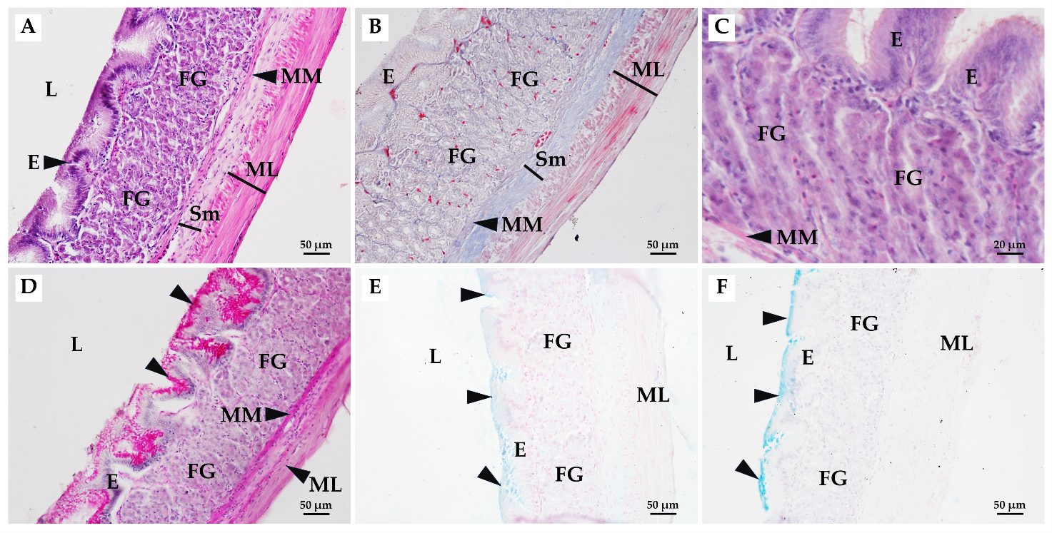
The striped tiger nandid fish, Pristolepis fasciata, is widely distributed in Thailand. However, its diet composition has intensively investigated, the histological profile remains interesting and challenging issues. Therefore, the objectives of this study are to describe the histological structure and histochemistry of the digestive tract of the striped tiger nandid fish. Fifteen adult fish were collected from Songkhla province, Thailand. The digestive tract was fixed in Bouin’s solution, and followed by basic paraffin techniques. The 5 µm sections were stained with hematoxylin and eosin (H&E), periodic acid Schiff’s (PAS), alcian blue (AB) pH 2.5 and pH 1.0, and Masson's trichrome (MT). The results revealed that the wall of the digestive tract comprised of four layers: mucosa, submucosa, muscularis, and serosa. The mucous and goblet cells in the esophagus exhibited positive staining with PAS, AB pH 2.5, and pH 1.0. The stomach was divided into three parts: cardiac, fundic, and pyloric stomach. The epithelium of the cardiac stomach showed positive staining with PAS and weak staining with AB pH 1.0. The fundic cells showed strong positive staining with PAS but weak staining with AB pH 2.5 and pH 1.0. Whereas the cells lining the fundic and cardiac glands showed positive staining with PAS. The pyloric epithelium revealed positive staining with PAS but did not contain gland. In the anterior, middle, and posterior intestines, glands were absent. Goblet cells in each portion exhibited intense labeling with both PAS and AB pH 2.5 and pH 1.0. The intestinal coefficient was 0.62±0.01, indicating an omnivorous fish.
Article Details

This work is licensed under a Creative Commons Attribution-NonCommercial-NoDerivatives 4.0 International License.
References
Cinar, K.; Nurgül, S. Histological and histochemical characterization of the mucosa of the digestive tract in flower fish (Pseudophoxinus antalyae). Anat. Histol. Embryol. 2006, 35, 147-151. https://doi.org/10.1111/j.1439-0264.2005.00629.x
Okuthe, G.E.; Bongile, B. Morphology, histology and histochemistry of the digestive tract of the banded tilapia, Tilapia sparrmanii (Perciformes: Cichlidae). Zoologia 2020, 37, 1-14.
Wainwright, P.C.; Richard, B.A. Predicting patterns of prey use from morphology of fishes. Environ. Biol. Fishes. 1995, 44, 97-113. https://doi.org/10.3897/zoologia.37.e51043
Al-Abdulhadi, H.A. Some comparative histological studies on alimentary tract of tilapia fish (Tilapia spilurus) and sea bream (Mylio cuvieri). Egypt. J. Aquat. Res. 2005, 31, 387-39.
Hernández, D.R.; Pérez Gianeselli, M.; Domitrovic, H.A. Morphology, histology and histochemistry of the digestive system of south American catfish (Rhamdia quelen). Int. J. Morphol. 2009, 27, 105-111. https://doi.org/10.4067/S0717-95022009000100019
Senarat, S.; Jes, K.; Wannee, J.; Niwat, K. Structural classifications in the digestive tract of short mackerel, Rastrelliger brachysoma (Bleeker, 1851) from upper gulf of Thailand. Songklanakarin J. Sci. Technol. 2015, 37, 561-567.
Borman, M.; Ismot, A.; Mohammad, K.; Md Rafiqun, N. Histo-morphology of the alimentary canal in two freshwater snakehead fish Channa punctata and Channa striata. J. Fish. 2015, 3, 297-300. https://doi.org/10.17017/j.fish.106
Kasozi, N.; Gerald, I.D.; Julius, M.; Charles, D.K.; Majid, K.; Akisoferi, O.W.; Godfrey, K.; Victoria, T.N. Histomorphological description of the digestive system of pebbly fish, Alestes baremoze (Joannis, 1835). Sci. World J. 2017, https://doi.org/10.1155/2017/8591249. https://doi.org/10.1155/2017/8591249
Kalhoro, H.; Shengli, T.; Lei, W.; Ying, H.; Josie, A.V.; Qingjun, S. Morphological study of the gastrointestinal tract of Larimichthys crocea (Acanthopterygii: Perciformes). Zoologia 2018, 35, 1-9. https://doi.org/10.3897/zoologia.35.e25171
Alabssawy, A.N.; Hassan, M.M.K.; Ahmed, A.G. Anatomical and histological adaptations of digestive tract in relation to food and feeding habits of lizardfish, Synodus variegatus (Lacepède, 1803). Egypt. J. Aquat. Res. 2019, 45, 159-165. https://doi.org/10.1016/j.ejar.2019.05.006
Santos, M.L.D.; Fábio, P.A.; Tiago, C.P.; José, E.S. Morphological, histological and histochemical analysis of the digestive tract of Trachelyopterus striatulus (Siluriformes: Auchenipteridae). Zoologia 2015, 32, 296-305. https://doi.org/10.1590/S1984-46702015000400005
Smith, L.S. Introduction to Fish Physiology, Argent Laboratories Press: Washington, DC., The United States of America, 1991.
Qu, M.; Shaoxiong, D.; Xiaojing, X.; Minghui, S.; Yingzhe, Y.; Yongquan, S. Ontogenetic development of the digestive system and growth in coral trout (Plectropomus leopardus). Aquaculture 2012, 334, 132-141. https://doi.org/10.1016/j.aquaculture.2011.12.046
Angelescu, V.; Franscisco, S.G. Adaptaciones del aparato digestivo al régimen alimenticio en algunos peces del Río Uruguay y Río de La Plata. Rev. Inst. Nac. Invest. Nat. 1949, 1, 161-281.
Rainboth, W.J. Fishes of the Cambodian Mekong. FAO Species Identification Field Guide for Fishery Purposes, FAO: Rome, Italy, 1996.
Sangpradub, N.; Chutima, H. Diet composition of Pristolepis fasciata (Bleeker, 1851) (Family Nandidae) and Puntius brevis (Bleeker, 1849) (Family Cyprinidae) in Kaeng Lawa, Thailand. Chiang Mai J. Sci. 2017, 44, 839-846.
Uk-katawewat, S. Illustration of Thai Fish and Aquatic animals, Kdurusapa Ladprao Printing Press: Bangkok, Thailand, 2002.
Bancroft, J.D.; Marilyn, G. Theory and practice of histological techniques, Churchill Livingstone: London, U.K., 2002.
Ghosh, S.K.; Padmanabha, C. Histological and histochemical characterization on stomach of Mystus cavasius (Hamilton), Oreochromis niloticus (Linnaeus) and Gudusia chapra (Hamilton): comparative study. The Journal of Basic and Applied Zoology 2015, 70, 16-24. https://doi.org/10.1016/j.jobaz.2015.04.002
Smith, B.J.; Smith, S.A.; Bundit, T.; Terry, A.L. Gross morphology and topography of the adult intestinal tract of the tilapian fish, Oreochromis niloticus L. Cells Tissues Organs 2000, 166, 294-303. https://doi.org/10.1159/000016743
Santos, M.D.; Lima, C.L.P.; Sandra, R.; Carvalho, B.S.M. Metazoan parasite fauna of Pimelodus maculatus La Cépède, 1803 (Siluriformes, Pimelodidae) from the Guandu river, Rio de Janeiro Rio State, Brazil. Acta Sci. Biol. Sci. 2007, 29, 101-107. https://doi.org/10.4025/actascibiolsci.v29i1.130
Vieira-Lopes, D.A.; Nadja, L.P.; Armando, S.; Adriana, V.; Francisco, G.A.; Iracema, D.G.; Aparecida, A.N. Immunohistochemical study of the digestive tract of Oligosarcus hepsetus. World J. Gastroenterol. 2013, 19, 1919-1929. https://doi.org/10.3748/wjg.v19.i12.1919
Nascimento, W.S.; Glaucia, M.M.S.; Lenilda, T.T.; Naisandra, B.S.; Sathyabama, C. Histology of the digestive tract of Anablepsoides urophthalmus from Brazilian oriental Amazonia. Journal of Aquaculture and Marine Biology 2018, 7, 39-42. https://doi.org/10.15406/jamb.2018.07.00181
Purushothaman, K.; Doreen, L.; Jolly, M.S.; Syed, M.S.; Declan, P.L.; Shubha, V.; László, O. Morpho- histological characterisation of the alimentary canal of an important food fish, Asian seabass (Lates calcarifer). PeerJ. 2016, DOI 10.7717/peerj.2377. https://doi.org/10.7717/peerj.2377
Dias, A.C.M.I.; Castelo, B.C.W.; Lopes, V.G. Estudo da dieta natural de peixes no reservatório de Ribeirão das Lajes, Rio de Janeiro, Brasil. Acta Sci. Biol. Sci. 2005, 7, 355-364. https://doi.org/10.4025/actascibiolsci.v27i4.1270
Machado, M.R.F.; Helena, O.S.; Valderes, L.S.; Alexandre, A.; Roberto, G.; André, D.B. Morphological and anatomical characterization of the digestive tract of Centropomus parallelus and C. undecimalis. Acta Sci. Biol. Sci. 2013, 35, 467-474.
Reifel, C.W.; Anthony, A.T. Structure and carbohydrate histochemistry of the stomach in eight species of teleosts. J. Morphol 1978, 158, 155-67. https://doi.org/10.1002/jmor.1051580203
Pedini, V.; Cecilia, D.; Parillo, F.; Paola, S. A lectin histochemical study of the oesophagus of Shi Drum. J. Fish. Biol. 2004, 64, 625-631. https://doi.org/10.1111/j.1095-8649.2004.00326.x
Sibbing, F.A.; Uribe, R. Regional specialisation in the oro-pharyngeal wall and food processing in carp (Cyprinus carpio L.). Neth. J. Zool. 1985, 35, 377-422. https://doi.org/10.1163/002829685X00280
Díaz, A.O.; Alocia, M.C.; Adriana, L.G. Glycoconjugates in the mucosa of the digestive tract of Cynoscion guatucupa: a histochemical study. Acta Histochem. 2008, 110, 76-85. https://doi.org/10.1016/j.acthis.2007.08.002
Canan, B.; Wallace, S.N.; Naisandra, B.S.; Sathyabama, C. Morphohistology of the digestive tract of the damsel fish Stegastes fuscus (Osteichthyes: Pomacentridae). The Scientific World Journal 2012, doi:10.1100/2012/787316. https://doi.org/10.1100/2012/787316
Rodrigues da Silva, M.; Maria, R.M.N.; Norma, S.H. Histology of the digestive tract of Satanoperca pappaterra (Osteichthyes, Cichlidae). Acta Sci. Biol. 2012, 34, 319-326. https://doi.org/10.4025/actascibiolsci.v34i3.8956
Cao, X.J.; Wang, W.M. Histology and mucin histochemistry of the digestive tract of yellow catfish, Pelteobagrus fulvidraco. Anat Histol Embryol. 2009, 38, 254-261. https://doi.org/10.1111/j.1439-0264.2009.00932.x
Eckert, R.; Randall, D.; Augustine, G. Animal Physiology; Mechanisms and Adaptations, WH Freeman and Company: New York, The United States, 1988.
Cardoso, N.N.; Enely, M.S.F.; Iracema, D.G.; Aparecida, A.N.; Armando, S.; Francisco, G.A. Histochemical and immunohistochemical study on endocrine cells (5HT, GAS, and SST) of the gastrointestinal tract of a teleost, the characin Astyanax bimaculatus. Acta Histochem. 2015, 117, 595-604. https://doi.org/10.1016/j.acthis.2015.05.007
Pessoa, E.K.R.; Naisandra, B.S.; Naithirithi, T.C.; Arrilton, A.S.; Sathyabama, C. Morfologia comparativa do trato digestorio dos peixes Hoplias malabaricus e Hypostomus pusarum do acude Marechal Dutra, Rio Grande do Norte, Brasil. Biota Amazôn. 2013, 3, 48-57. https://doi.org/10.18561/2179-5746/biotaamazonia.v3n1p48-57
Andrade, I.M.; Juliana, P.G.; Matheus, M.R.; Renata, B.M. Morphology of the digestive tract of the whitemouth croaker Micropogonias furnieri (Desmarest, 1823) (Perciformes: Sciaenidae). Acta Zool. 2016, doi: 10.1111/azo.12156. https://doi.org/10.1111/azo.12156
Legler, J.M. Morphology and physiology of the Chelonia. Australian Biological Recourses Study, Caberra 1993, 108-119.
Domeneghini, C.; Silvana, A.; Giuseppe, R.; Giampaolo, B.; Veggetti A. Histochemical analysis of glycoconjugate secretion in the alimentary canal of Anguilla anguilla L. Acta Histochem. 2005, 106, 477-87. https://doi.org/10.1016/j.acthis.2004.07.007
Rotta, M.A. Aspectos gerais da fisiologia e estrutura do sistema digestivo dos peixes relacionados à piscicultura, Corumbá Embrapa: Brazil, 2003.
Ringø, E.; Reidar, M.; Terry, M.M.; Rolf, E.O. Bacterial translocation and pathogenesis in the digestive tract of larvae and fry. Aquaculture 2007, 268, 251-264. https://doi.org/10.1016/j.aquaculture.2007.04.047
Carrassón, M.; Grau, A.; Dopazo, L.R.; Selena, C. A histological, histochemical and ultrastructural study of the digestive tract of Dentex dentex (Pisces, Sparidae). Histol. Histopathol. 2006, 21, 579-593.
Anderson, T.A. Histological and cytological structure of the gastrointestinal tract of the luderick, Girella tricuspidata (Pisces, Kyphosidae), in relation to diet. J. Morphol. 1986, 190, 109-119. https://doi.org/10.1002/jmor.1051900110
Clarke, A.J.; Witcomb, D.M. A study of the histology and morphology of the digestive tract of the common eel (Anguilla anguilla). J. Fish. Biol. 1980, 16, 159-170. https://doi.org/10.1111/j.1095-8649.1980.tb03695.x
Gartner, L.P.; Hiatt, J.L.; De Souza, L.F.; Sales, Md.G.F. Atlas colorido de histologia, Guanabara Koogan LTDA: Rio de Janeiro, Brazil, 2002.
