
Histology and Histochemistry of the Digestive Tract of Transverse-bar Barb, Hampala macrolepidota Kuhl & Van Hasselt, 1823
Article Sidebar
Main Article Content
Abstract
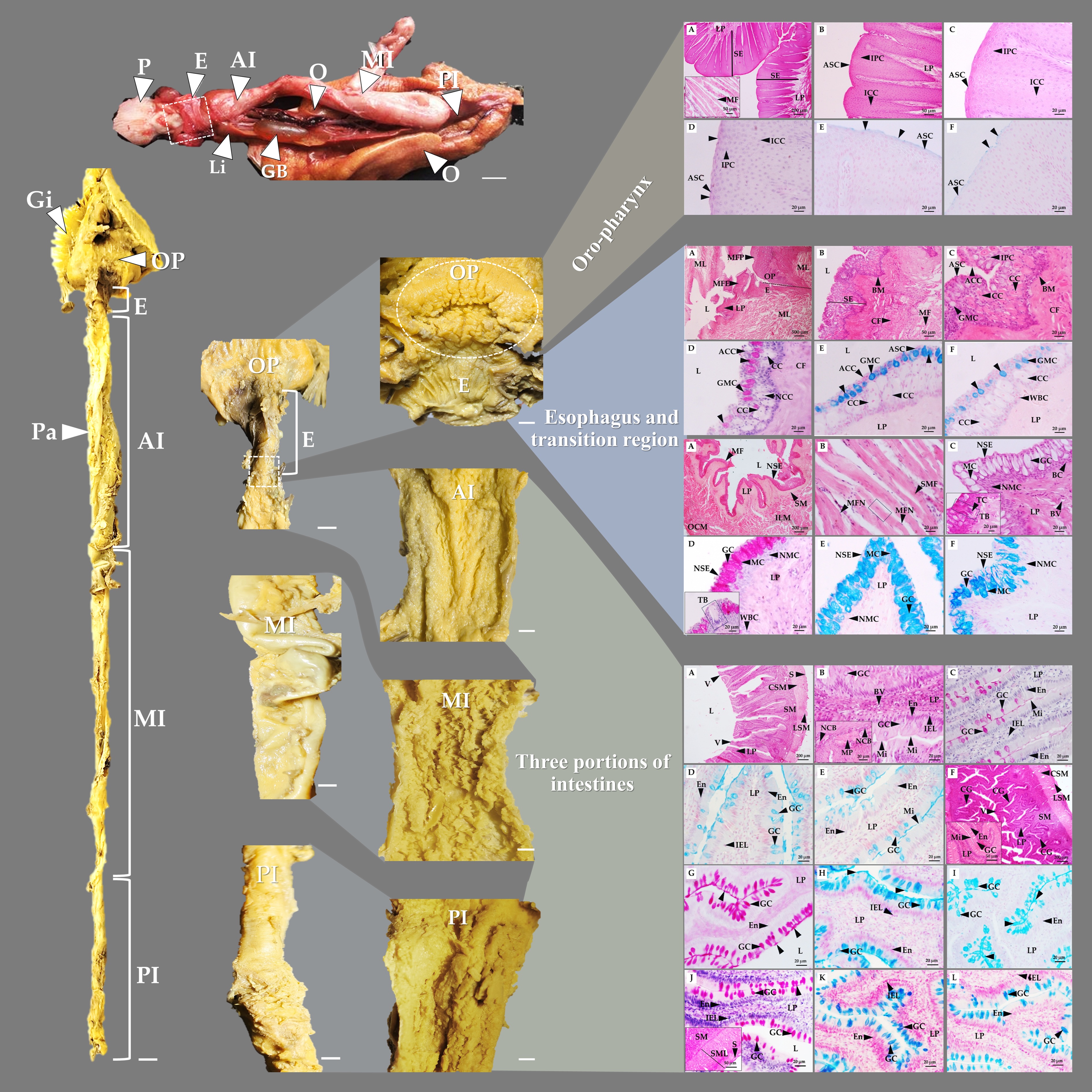
Hampala macrolepidota is an important local fishery species; the information on its anatomy and microanatomy is scarce. Therefore, the histology and histochemistry of its digestive tract were systematically investigated. The digestive tracts of adult fish were removed and processed using the paraffin technique. Sections of 5 µm thickness were stained with hematoxylin and eosin (H&E), periodic acid-Schiff’s (PAS), and alcian blue (AB), pH 2.5 and pH 1.0. Its digestive tract consisted of the oropharynx, esophagus, and intestine, with no stomach observed. The stratified epithelium of the oropharynx was weakly positive-stained with PAS and AB at pH 2.5 and pH 1.0. In contrast, in the esophagus, mucous and goblet cells were strongly positively stained with all stains, indicating the secretion of neutral mucin and carboxylated and sulfated acid mucins. The intestine can be divided into three regions: anterior, middle, and posterior. Meanwhile, the enterocytes were positively stained with PAS; however, the goblet cells were positively stained with PAS at pH 2.5 and pH 1.0. The goblet cells secreted neutral mucins, as well as carboxylated and sulfated acidic mucins. The middle intestine showed crypt-like glands and the highest histometric values, indicating that it served as the main site of digestion and nutrient absorption, despite the absence of a stomach. The muscular layer varied regionally, with skeletal muscle in the oropharynx and esophagus and smooth muscle in the intestine. The intestinal coefficient was 0.69, indicating that it was a carnivorous fish. These findings provided fundamental data relevant to aquaculture, ecology, and comparative vertebrate anatomy.
Article Details

This work is licensed under a Creative Commons Attribution-NonCommercial-NoDerivatives 4.0 International License.
References
Al Abdulhadi, H. A. Some comparative histological studies on alimentary tract of tilapia fish (Tilapia spilurus) and sea bream (Mylio cuvieri). Egypt. J. Aquat. Res. 2005, 31(1), 387–398.
Fagundes, K. R. C.; Rotundo, M. M.; Mari, R. B. Morphological and histochemical characterization of the digestive tract of the puffer fish Sphoeroides testudineus (Linnaeus, 1758) (Tetraodontiformes: Tetraodontidae). An. Acad. Bras. Cienc. 2016, 88(3), 1615–1624. https://doi.org/10.1590/0001-3765201620150167
Çinar, K.; Senol, N. Histological and histochemical characterization of the mucosa of the digestive tract in flower fish (Pseudophoxinus antalyae). Anat. Histol. Embryol. 2006, 35(3), 147–151. https://doi.org/10.1111/j.1439-0264.2005.00629.x
Okuthe, G. E.; Bhomela, B. Morphology, histology and histochemistry of the digestive tract of the banded tilapia, Tilapia sparrmanii (Perciformes: Cichlidae). Zoologia 2020, 37, e51043. https://doi.org/10.3897/zoologia.37.e51043
Dabrowski, K. R.; Portella, M. C. Feeding plasticity and nutritional physiology in tropical fishes. Fish Physiol. 2005, 21, 155–224. https://doi.org/10.1097/01.ruq.0000177221.14263.45
Pavelin, T.; Kević, N.; Restović, I.; Bočina, I. Histological and biochemical features of the digestive system in the cage-reared gilthead sea bream (Sparus aurata). Int. J. Biol. Res. 2018, 2(1), 51–56. https://doi.org/10.18689/ijbr-1000108
Genten, F.; Terwinghe, E.; Danguy, A. Atlas of Fish Histology, 1st ed.; CRC Press: Boca Raton, FL, 2009.
Senarat, S.; Yenchum, W.; Poolprasert, P. Histological study of the intestine of Stoliczkae's barb Puntius stoliczkanus (Day, 1871) (Cypriniformes: Cyprinidae). Kasetsart J. Nat. Sci. 2013, 47, 247–251.
Aruho, C.; Namulawa, V.; Kato, C. D.; Kisekka, M.; Rutaisire, J.; Bugenyi, F. Histo-morphological description of the digestive system of the rippon barbel Barbus altianalis (Boulenger, 1900): A potential species for culture. Uganda J. Agric. Sci. 2016, 17(2), 197–217. https://doi.org/10.4314/ujas.v17i2.6
Bhat, M. Y.; Channa, A.; Paray, B. A.; Al-Sadoon, M. K.; Rather, I. A. Morphological study of the gastrointestinal tract of the snow trout, Schizothorax esocinus (Actinopterygii: Cypriniformes). Zoologia 2019, 36, e31791. https://doi.org/10.3897/zoologia.36.e31791
Gonçalves, M.; Lopes, C.; Silva, P. Comparative histological description of the intestine in platyfish (Xiphophorus maculatus) and swordtail fish (Xiphophorus helleri). Tissue Cell 2024, 87, 102306. https://doi.org/10.1016/j.tice.2024.102306
Kalhoro, H.; Tong, S.; Wang, L.; Hua, Y.; Volatiana, J. A.; Shao, Q. Morphological study of the gastrointestinal tract of Larimichthys crocea (Acanthopterygii: Perciformes). Zoologia 2018, 35, 1–9. https://doi.org/10.3897/zoologia.35.e25171
Raji, A. R.; Norouzi, E. Histological and histochemical study on the alimentary canal in walking catfish (Clarias batrachus) and piranha (Serrasalmus nattereri). Iran. J. Vet. Res. 2010, 11(3), 255–261.
Shalaby, W. Comparative morphological and histological studies on the adaptation of esophagus and stomach to the feeding habits in some coral reef fishes at Hurghada, Red Sea, Egypt. Egypt. J. Aquat. Res. 2020, 24 (5), 289–306. https://doi.org/10.21608/ejabf.2020.105059
Evans, D. H.; Claiborne, J. B. The Physiology of Fishes, 3rd ed.; CRC Press: Boca Raton, FL, 2006.
Ortiz-Ruiz, M.; López Flórez, C.; Castro Rebolledo, M. I.; Baldisserotto, B.; Gómez Ramírez, E. Anatomy, histology and ultrastructure of the digestive tract in Andean fish (Trichomycterus bogotensis) and ecological implications. Zoomorphology 2024, 143, 433–441. https://doi.org/10.1007/s00435-023-00634-3
Xiong, D.; Zhang, L.; Yu, H.; Xie, C. A study of morphology and histology of the alimentary tract of Glyptosternum maculatum (Sisoridae, Siluriformes). Acta Zool. 2011, 92, 161–169. https://doi.org/10.1111/j.1463-6395.2010.00458.x
Cyrino, J. P. E.; Bureau, D. P.; Kapoor, B. G. Feeding and Digestive Functions of Fish, 1st ed.; CRC Press: Boca Raton, FL, 2008.
Yuge, S.; Yamagami, S.; Inoue, K.; Suzuki, N.; Takei, Y. Identification of two functional guanylin receptors in eel: Multiple hormone-receptor system for osmoregulation in fish intestine and kidney. Gen. Comp. Endocrinol. 2006, 149, 10–20. https://doi.org/10.1016/j.ygcen.2006.04.012
Grosell, M.; Taylor, J. R. Intestinal anion exchange in teleost water balance. Comp. Biochem. Physiol. A Mol. Integr. Physiol. 2007, 148(1), 14–22. https://doi.org/10.1016/j.cbpa.2006.10.017
Brandtzaeg, P. Mucosal immunity: Induction, dissemination, and effector functions. Scand. J. Immunol. 2009, 70(6), 505–515. https://doi.org/10.1111/j.1365-3083.2009.02319.x
Vieira-Lopes, D. A.; Pinheiro, N. L.; Sales, A.; Ventura, A.; Araújo, F. G.; Gomes, I. D.; Nascimento, A. A. Immunohistochemical study of the digestive tract of Oligosarcus hepsetus. World J. Gastroenterol. 2013, 19, 1919–1929. https://doi.org/10.3748/wjg.v19.i12.1919
Purushothaman, K.; Lau, D.; Saju, J. M.; Musthaq, S.; Lunny, D. P.; Vij, S.; Orbán, L. Morpho-histological characterisation of the alimentary canal of an important food fish, Asian seabass (Lates calcarifer). PeerJ 2016, 4, e2377. https://doi.org/10.7717/peerj.2377
Na Lampang, P.; Palasai, A.; Senarat, S.; Jiraungkoorskul, W.; Kettratad, J. Observation of gut content and morpho-histology of the digestive system in Pisodonophis boro (Hamilton, 1822) from Pranburi River Estuary, Thailand. Songklanakarin J. Sci. Technol. 2021, 43(2), 496–504.
Kasozi, N.; Iwe, G. D.; Langi, S.; Namulawa, V. T.; Walakira, J. Histological features of the gastrointestinal tract of elongate tigerfish, Hydrocynus forskahlii (Cuvier, 1819), from Lake Albert. J. Basic Appl. Zool. 2024, 85, 11. https://doi.org/10.1186/s41936-024-00364-y
Currie, S.; Evans, D. H. The Physiology of Fishes, 5th ed.; CRC Press: Boca Raton, FL, 2021.
Ghosh, S. K.; Chakrabarti, P. Histological and histochemical characterization on stomach of Mystus cavasius, Oreochromis niloticus and Gudusia chapra: Comparative study. J. Basic Appl. Zool. 2015, 70, 16–24. https://doi.org/10.1016/j.jobaz.2015.04.002
Guillard, J.; Tessier, A.; Beaune, D.; Kue, K.; Cottet, M.; Chanudet, V.; Descloux, S.; Panfili, J. Life History Traits and Exploitation of Hampala macrolepidota (Cyprinidae) in a Subtropical Reservoir (Lao PDR). Cybium 2019, 43(4), 351–365.
Titin, H.; Yustiatia, A.; Nurhayatia, A. Growth Patterns and Reproduction of Hampala macrolepidota in Jatigede Reservoir, West Java, Indonesia. InaJL 2022, 3(2), 66–75. https://doi.org/10.51264/inajl.v3i2.33
Ahmad, A. B. Hampala macrolepidota. The IUCN Red List of Threatened Species 2019, e.T181255A1714119. https://doi.org/10.2305/IUCN.UK.2019-2.RLTS.T181255A1714119.en (accessed October 31, 2025).
Risdawati, R.; Dahelmi, D.; Nurdin, J.; Syandri, H. Bioecological Aspects of Hampala macrolepidota in Lake Singkarak, West Sumatera, Indonesia. AACL Bioflux 2020, 13(2), 893–901.
Liu, M. D.; Huang, F. J.; Zhu, J. Z.; Liu, R. C.; Liu, S. P. Reproductive Biology of Hampala macrolepidota. Chin. J. Zool. 2015, 50(3), 405–414.
Bancroft, J. D.; Gamble, M. Theory and Practice of Histological Techniques; Churchill Livingstone: London, U.K., 2002.
Bočina, I.; Šantić, Ž.; Restović, I.; Topić, S. Histology of the Digestive System of the Garfish Belone belone. Eur. Zool. J. 2017, 81(1), 89–95.
de Oliveira Ribeiro, C. A.; Fanta, E. Microscopic Morphology and Histochemistry of the Digestive System of Trichomycterus brasiliensis. Rev. Bras. Zool. 2000, 17(4), 953–971. https://doi.org/10.1590/S0101-81752000000400007
Gómez-Ramírez, E.; Tovar, O.; Bulla, M. J. O.; Hurtado, H. Estudio Histológico del Tracto Digestivo del Pez Ariopsis seemanni. Rev. Fac. Cienc. Básicas 2010, 6(2), 216–225.
Beltran, L. G.; Santana, V. D.; Verdugo, M. H.; Gómez-Ramírez, E.; Hurtado-Giraldo, H. Descripción Anatómica e Histológica del Tracto Digestivo de Pimelodus blochii. Orinoquia 2013, 17(1), 102–110. https://doi.org/10.22579/20112629.55
Olaya, C. M.; Ovalle, C. H.; Gómez, E.; Rodríguez, D.; Caldas, M. L.; Hurtado, H. Histología y Morfometría del Sistema Digestivo de Pimelodus pictus. Rev. Fac. Med. Vet. Zootec. 2007, 54, 311–323.
Elliott, D. G. Integumentary System; Academic Press: United Kingdom, 2000. https://doi.org/10.1016/B978-012529650-2/50008-1
Mello, G. C. G.; Santos, M. L.; Arantes, F.; Pessali, T. C. Morphological Characterisation of the Digestive Tract of Lophiosilurus alexandri. Acta Zool. 2017, 1–10.
Chivers, D. P.; Wisenden, B. D.; Hindman, C. J.; Michalak, T. A.; Kusch, C.; Kaminskyj, S. G. W.; Jack, K. L.; Ferrari, M. C. O.; Pollock, R. J.; Halbgewachs, C. F.; Pollock, M. S.; Alemadi, S.; James, C. T.; Savaloja, R. K.; Goater, C. P.; Corwin, A.; Mirza, R. S.; Kiesecker, J. M.; Brown, G. E.; Adrian, J. C.; Krone, P. H.; Blaustein, A. R.; Mathis, A. Epidermal Alarm Substance Cells of Fishes Maintained by Non-Alarm Functions. Proc. R. Soc. B 2007, 1–9.
Nazlić, M.; Paladini, A.; Bočina, I. Histology of the Digestive System of Scorpaena porcus. Acta Adriat. 2014, 55(1), 65–74.
Machado, M. R. F.; de Oliveira Souza, H.; de Souza, V. L.; de Azevedo, A.; Goitein, R.; Nobre, A. D. Morphological and Anatomical Characterization of the Digestive Tract of Centropomus parallelus and C. undecimalis. Acta Sci. 2013, 35(4), 467–474. https://doi.org/10.4025/actascibiolsci.v35i4.14352
Pewhom, A.; Vanikasampanna, A. Histological Structure and Histochemistry of the Digestive Tract of Pristolepis fasciata. ASEAN J. Sci. Technol. Rep. 2024, 27(4). https://doi.org/10.55164/ajstr.v27i4.253044
Morais, S. The Physiology of Taste in Fish: Potential Implications for Feeding Stimulation and Gut Chemical Sensing. Rev. Fish. Sci. Aquac. 2017, 1–17.
Pedini, V.; Dall'Aglio, C.; Parillo, F.; Scocco, P. A Lectin Histochemical Study of the Oesophagus of Shidrum. J. Fish Biol. 2004, 64, 625–631. https://doi.org/10.1111/j.1095-8649.2004.00326.x
Abd, E. A.; Hafez, E.; Mokhtar, D.; Abou-Elhamd, A. S. Comparative Histomorphological Studies on Oesophagus of Catfish and Grass Carp. J. Histol. 2013, 1–10. https://doi.org/10.1155/2013/858674
Mittal, R. K.; Holloway, R. H.; Penagini, R.; Blackshaw, L. A.; Dent, J. Transient Lower Esophageal Sphincter Relaxation. Gastroenterology 1995, 109(2), 601–610. https://doi.org/10.1016/0016-5085(95)90351-8
Wilson, J. M.; Castro, L. F. C. Morphological Diversity of the Gastrointestinal Tract in Fishes. In Fish Physiology: The Multifunctional Gut of Fish; Grosell, M.; Farrell, A. P.; Brauner, C. J., Eds.; Academic Press: United States, 2011; pp 1–55.
Sangsawang, A.; Vaniksampanna, A.; Intachai, S.; Pewhom, A. Histological and Histochemical Characteristics of the Digestive Tract of Mastacembelus favus. Trends Sci. 2025, 22(11), 10687. https://doi.org/10.48048/tis.2025.10687
Bebić, M.; Kević, N.; Restović, I.; Šantić, M.; Bočina, I. Histological and Histochemical Studies of Digestive System in Lepidorhombus whiffiagonis. Iran. J. Ichthyol. 2020, 7, 125–135.
Grosell, M.; Farrell, A. P.; Brauner, C. J. The Multifunctional Gut of Fish; Academic Press: United States, 2011.
Scocco, R.; Pedini, V. Histochemical Characterisation of Complex Carbohydrates Expressed in the Alimentary Tract of Chickens. Vet. J. 2010, 185, 228–230. https://doi.org/10.1016/j.tvjl.2009.04.015
Day, R. D.; German, D. P.; Manjakasy, J. M.; Farr, I.; Hansen, M. J.; Tibbetts, I. R. Enzymatic Digestion in Stomachless Fishes. J. Comp. Physiol. B 2011, 181, 603–613.
Canan, B.; do Nascimento, W. S.; da Silva, N. B.; Chellappa, S. Morphohistology of the Digestive Tract of Stegastes fuscus. Sci. World J. 2012, 787316, 1–9. https://doi.org/10.1100/2012/787316
Carrassón, M.; Grau, A.; Dopazo, L. R.; Crespo, S. A Histological, Histochemical and Ultrastructural Study of the Digestive Tract of Dentex dentex. Histol. Histopathol. 2006, 21, 579–593.
Quintana-Hayashi, M. P.; Mahu, M.; De Pauw, N.; Boyen, F.; Pasmans, F.; Martel, A.; Premaratne, P.; Fernandez, H. R.; Teymournejad, O.; Maele, L. V.; Haesebrouck, F.; Lindén, S. K. Binding of Brachyspira hyodysenteriae to Porcine Colonic Mucins. Infect. Immun. 2015, 83(4), 1610–1619. https://doi.org/10.1128/IAI.03073-14
Lu, F.; Li, Y.; Wang, X.; Hu, X.; Liao, X.; Zhang, Y. Early-Life Polyphenol Intake Promotes Akkermansia Growth. Food Res. Int. 2021, 149, 110648. https://doi.org/10.1016/j.foodres.2021.110648
García, A.; Hernández, J.; Pardo, S. C. Descripción Morfológica del Tubo Digestivo de Juveniles de Salminus affinis. Acta Biol. Colomb. 2008, 13, 99–112.
