
Chronic Heat Stress-Induced Alterations in Hormonal Profile (Corticosterone, T3, T4, TSH) and Oxidative Stress Markers (MDA, GSH, GPx) in Laboratory Rats
Article Sidebar
Main Article Content
Abstract
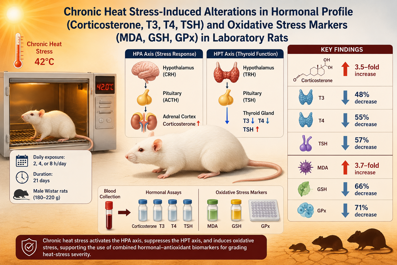
Chronic heat stress disrupts neuroendocrine balance and promotes oxidative damage, yet the duration-dependence of simultaneous hypothalamic-pituitary-adrenal (HPA) and hypothalamic-pituitary-thyroid (HPT) axis responses and antioxidant status remains poorly defined. This study quantified the effects of daily heat exposure (2, 4, or 8 h/day at 42°C for 21 days) on serum corticosterone, triiodothyronine (T3), thyroxine (T4), and thyroid-stimulating hormone (TSH), and on hepatic oxidative markers malondialdehyde (MDA), reduced glutathione (GSH), and glutathione peroxidase (GPx) in adult male Wistar rats. Thirty-two rats (180–220 g) were randomized into four groups (n = 8): control and three heat-stressed groups (HS-2, HS-4, HS-8). ELISA quantified hormones, and oxidative markers were measured spectrophotometrically; data were analyzed by one-way ANOVA with Tukey HSD and Pearson correlation. Heat stress produced duration-dependent endocrine and oxidative changes (p < 0.001). Corticosterone rose 3.5-fold, from 45.2 ± 4.1 to 158.7 ± 13.2 ng/mL in HS-8, whereas T3, T4, and TSH fell by 48%, 55%, and 57%, respectively. MDA increased 3.7-fold (2.14 ± 0.18 to 7.88 ± 0.61 nmol/mL), and GSH and GPx declined by 66% and 71%. Corticosterone correlated positively with MDA (r = +0.892) and negatively with GSH (r = −0.874); T3 correlated negatively with MDA (r = −0.861) and T4 positively with GPx (r = +0.847). Chronic heat stress, therefore, causes duration-dependent HPA-axis activation, HPT-axis suppression, and antioxidant depletion, and the tight hormone-redox coupling supports the use of combined hormonal-antioxidant biomarker panels for grading heat-stress severity.
Article Details

This work is licensed under a Creative Commons Attribution-NonCommercial-NoDerivatives 4.0 International License.
References
Bouchama, A.; Knochel, J. P. Heat stroke. N. Engl. J. Med. 2002, 346, 1978–1988. https://doi.org/10.1056/NEJMra011089
Speakman, J. R.; Keijer, J. Not so hot: optimal housing temperatures for mice to mimic the thermal environment of humans. Mol. Metab. 2013, 2, 5–9. https://doi.org/10.1016/j.molmet.2012.10.002
Dhabhar, F. S.; McEwen, B. S.; Spencer, R. L. Corticosterone regulation of hippocampal glucocorticoid receptor binding is cell type-specific. Brain Res. 1994, 648, 247–261. https://doi.org/10.1016/0006-8993(94)91124-5
McEwen, B. S. Physiology and neurobiology of stress and adaptation: central role of the brain. Physiol. Rev. 2007, 87, 873–904. https://doi.org/10.1152/physrev.00041.2006
Helmreich, D. L.; Parfitt, D. B.; Lu, X. Y.; Akil, H.; Watson, S. J. Relation between the hypothalamic-pituitary-thyroid (HPT) axis and the hypothalamic-pituitary-adrenal (HPA) axis during repeated stress. Neuroendocrinology 2006, 81, 183–192. https://doi.org/10.1159/000087001
Halliwell, B.; Gutteridge, J. M. C. Free Radicals in Biology and Medicine, 5th ed.; Oxford University Press: Oxford, UK, 2015. https://doi.org/10.1093/acprof:oso/9780198717478.001.0001
Sies, H. Oxidative stress: from basic research to clinical application. Am. J. Med. 1991, 91, 31S–38S. https://doi.org/10.1016/0002-9343(91)90281-2
Majhwol, E. M. Chia Seed Extract Attenuates Streptozotocin-Induced Pancreatic Beta-Cell Dysfunction in Diabetic Rats: Evidence from Functional and Oxidative Stress Biomarkers. ASEAN J. Sci. Tech. Report. 2026, 29(5), e263641. https://doi.org/10.55164/ajstr.v29i5.263641
Jawad, Z. H.; Salman, H. A. Gastroprotective Effects of Purslane (Portulaca oleracea L.) Ethanolic Extract Against Aspirin-Induced Gastric Ulcer: Modulation of Oxidative Stress, Inflammatory Mediators, and EGFR Signaling in Rats. ASEAN J. Sci. Tech. Report. 2026, 29(5), e263937. https://doi.org/10.55164/ajstr.v29i5.263937
Zahwar, A. T.; Sabour, A. N. Berberine Ameliorates Methionine-Induced Hyperhomocysteinemia and Biochemical Alterations in Male Rats. ASEAN J. Sci. Tech. Report. 2026, 29(3), e262094. https://doi.org/10.55164/ajstr.v29i3.262094
Gupta, A.; Yadav, R.; Samisha; Sawale, J.; Yadav, S. Sage and Rosemary Extract Gel: Anti-Aging Efficacy in D-galactose-Induced Skin Aging Model. ASEAN J. Sci. Tech. Report. 2024, 27(6), e254020. https://doi.org/10.55164/ajstr.v27i6.254020
Al-Musawi, S. L.; Al-Shamsi, M. A.; Hassan, N. H. Heat stress-induced neuroendocrine disruption and antioxidant imbalance in male Wistar rats: a dose-response analysis. Iraqi J. Vet. Sci. 2022, 36, 1015–1024. https://doi.org/10.33899/ijvs.2022.133120.2233
Mohammed, A. K.; Hassan, Y. I.; Saleh, R. A. Duration-dependent effects of chronic heat exposure on thyroid axis hormones and hepatic oxidative stress in laboratory rats. Comp. Clin. Pathol. 2023, 32, 477–486. https://doi.org/10.1007/s00580-023-03452-8
Zhang, F.; Dou, J.; Zhao, X.; Luo, H.; Ma, L.; Wang, L.; Wang, Y. Corticosterone and catecholamine responses in heat-stressed rats: adrenal transcriptomic and proteomic profiling. Animals 2023, 13, 1618. https://doi.org/10.3390/ani13101618
Ohkawa, H.; Ohishi, N.; Yagi, K. Assay for lipid peroxides in animal tissues by thiobarbituric acid reaction. Anal. Biochem. 1979, 95, 351–358. https://doi.org/10.1016/0003-2697(79)90738-3
Ellman, G. L. Tissue sulfhydryl groups. Arch. Biochem. Biophys. 1959, 82, 70–77. https://doi.org/10.1016/0003-9861(59)90090-6
Flohé, L.; Günzler, W. A. Assays of glutathione peroxidase. Methods Enzymol. 1984, 105, 114–121. https://doi.org/10.1016/S0076-6879(84)05015-1
McEwen, B. S.; Wingfield, J. C. The concept of allostasis in biology and biomedicine. Horm. Behav. 2003, 43, 2–15. https://doi.org/10.1016/S0018-506X(02)00024-7
Dhabhar, F. S. Effects of stress on immune function: the good, the bad, and the beautiful. Immunol. Res. 2014, 58, 193–210. https://doi.org/10.1007/s12026-014-8517-0
Sapolsky, R. M.; Romero, L. M.; Munck, A. U. How do glucocorticoids influence stress responses? Integrating permissive, suppressive, stimulatory, and preparative actions. Endocr. Rev. 2000, 21, 55–89. https://doi.org/10.1210/edrv.21.1.0389
Kalsbeek, A.; Fliers, E.; Franke, A. N.; Wortel, J.; Buijs, R. M. Functional connections between the suprachiasmatic nucleus and the thyroid gland as revealed by lesioning and viral tracing techniques in the rat. Endocrinology 2000, 141, 3832–3841. https://doi.org/10.1210/endo.141.10.7709
Wang, X.; Huang, Z.; Li, H.; Zhu, J.; Chen, T. Chronic heat stress modulates hypothalamic-pituitary-adrenal and thyroid axes in rats via glucocorticoid-TRH crosstalk: evidence from in vivo and ex vivo approaches. Front. Endocrinol. 2024, 15, 1342892. https://doi.org/10.3389/fendo.2024.1342892
Danforth, E., Jr.; Burger, A. The role of thyroid hormones in the control of energy expenditure. Clin. Endocrinol. Metab. 1984, 13, 581–595. https://doi.org/10.1016/S0300-595X(84)80039-0
Zhao, J.; Li, S.; Lin, Q.; Chen, G. Lipid peroxidation and antioxidant status in heat-stressed laboratory animals: a systematic review and meta-analysis. Oxid. Med. Cell. Longev. 2022, 2022, 9843750. https://doi.org/10.1155/2022/9843750
Dokladny, K.; Myers, O. B.; Bharat, A. M. Heat shock response and autophagy: cooperation and interconnection. Autophagy 2015, 11, 702–716. https://doi.org/10.1080/15548627.2015.1009776
Bouchama, A.; Abuyassin, B.; Lehe, C.; Laitano, O.; Jay, O.; Leon, L. R.; Roberts, W. O. Classic and exertional heatstroke. Nat. Rev. Dis. Primers 2022, 8, 8. https://doi.org/10.1038/s41572-021-00334-6
Aluigi, M. G.; De Flora, A.; Traverso, N. Glutathione as a key mediator in heat shock response: mechanisms and therapeutic implications. Free Radic. Biol. Med. 2023, 198, 45–58. https://doi.org/10.1016/j.freeradbiomed.2022.11.012
Manoli, I.; Alesci, S.; Blackman, M. R.; Su, Y. A.; Rennert, O. M.; Chrousos, G. P. Mitochondria as key components of the stress response. Trends Endocrinol. Metab. 2007, 18, 190–198. https://doi.org/10.1016/j.tem.2007.04.004
Venditti, P.; Di Meo, S.; De Rosa, G. Role of thyroid state in the vulnerability to oxidative stress induced by metabolic exhaustion in rat liver. Free Radic. Res. 2015, 49, 436–446. https://doi.org/10.3109/10715762.2015.1014961
